 Fitness, Nutrition, Health and Sports Blog In the HSNstore Blog you will find tips about Fitness, sport in general, nutrition, and health – HSNstore.com

We’re going to analyse the relationship between the amount of repetitions executed and hypertrophy based on the loading and activation of the corresponding signaling pathways
In December 2017, I published an article on this blog entitled Hybrid Training: Get Greater Muscle Hypertrophy.
In it, I explained the differences between a strength training routine oriented towards the improvement of your repetition maximum (1RM) and one orientated towards the development of musculoskeletal hypertrophy.
I mentioned, without going into too much detail, the role of mechanical stress and metabolic stress on signalling that can generate hypertrophy, and concluding that combining different training programmes with high and low loads intra- or inter-session was possibly the best approach to develop hypertrophy.
Today, I’m going to explain why this happens at a biomolecular level. I won’t go over the basic concepts we already looked at in the last article, so I recommend giving it a read first.
OK, let’s get to it!
Continuous Hypertrophy
We know that the equalising element of training aimed at increasing muscle hypertrophy is the intensity
This is not to be confused with external loading, rather it’s an internal stress scheme (check out how to measure effort to get a better understanding of it).
With this in mind, we have already moved away from the classic pattern of strict repetition regimes where:
- 1-5 repetitions are used to improve strength,
- 6-12 for hypertrophy, and
- +15 for strength-resistance.
This is because as we approach muscle failure we’ll reach the same amount of effective repetitions in both cases, which is the moment when the bulk of the motor units present in the stimulated musculature are recruited.
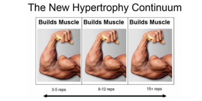
Hypertrophy continuum model (Schoenfeld, 2018)
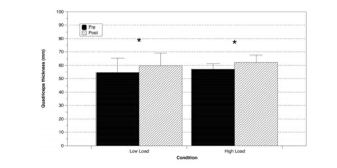
Changes in the cross-sectional area (mm) pre-post intervention in the group subjected to low and high loads (Schoenfeld, Peterson, Ogborn, Contreras & Sonmez, 2015)
Do the amount of repetitions matter?
Although a moderate range, such as the classic 8-12, is the most efficient in terms of benefit:time invested, the entire repetition range generates a similar hypertrophy.
Until recently we didn’t know exactly the ways in which this happened, or even whether the range of repetitions at the same effective repetitions and effort ratio was in fact completely irrelevant.
Study
Lysenko, Popov, Vepkhadze, Sharova and Vinogradova (2019) published a study clarifying the responses in autocrine and endocrine signalling that generate different loads (high and moderate) in trained subjects.
In the study, a total of 8 trained subjects carried out 4 sets of unilateral leg presses at maximum volitional effort using moderate (65%) and high (85% 1RM) load for the contralateral leg. The study showed a similar increase in muscle protein synthesis in both groups.
We know that acute protein synthesis is a reliable, though not absolute, indicator of long-term cross-sectional area increase (Mitchell et al. 2015); but as the study was developed in a single session, they’ve not been able to control the structural changes generated by both protocols on the subjects.
It’s important to note that the workload of the moderate load group is 32% higher than that of the high load group.
Results
Something that seems to indicate that, as the protein synthesis response is similar in both groups, the absolute volume of work, even the tonnage (load*repetitions) is a factor of little relevance as the volume is equalised when the effort ratio is maximal in both groups.
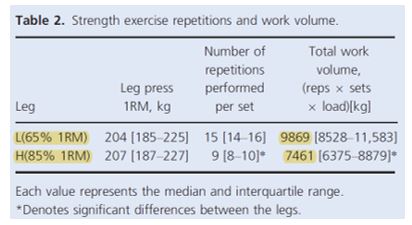
Volume of work in the modelled load group (L) and high load group (H). (Lysenko, 2019)
Loads and activation of signalling routes
Here an image to help explain, looking at the signalling pathways activated by each load protocol:
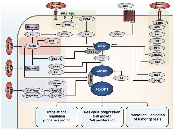
Signalling mechanisms that mediate cell growth and proliferation
Biopsies showed that moderate loads more significantly activated the mTORC1 pathway, presumably by a mechanism dependent on the high work volume, moderate loads increased phosphorylation of P70S6K, a river substrate below the consequent activation of the AKT/mTORC1 pathway that regulates ribosome synthesis (protein producers), in the myocyte (muscle cell).
Therefore, its activation, linked to a blockade of the phosphorylation of 4E-BP1, increases the expression of the RAPTOR subunit of mTORC1, which, in turn, through moderate loads, phosphoryl threonine46 is sufficient to bind slightly to eIF4E, avoiding the significant binding of eIF4E:4E-BP1, meaning it doesn’t interfere with its activity that positively regulates cell growth.
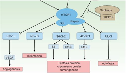
Influence of Mtorc1 subunit activation on protein synthesis
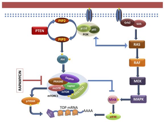
Mechanisms of ribosomal protein translation regulation mediated by P70S6K and eIF4E
High loads activated dephosphorylated eEF2, a key translation and elongation factor in protein synthesis processes during gene translation (moving mRNA to the ribosome and in the formation of polypeptides by tRNA)
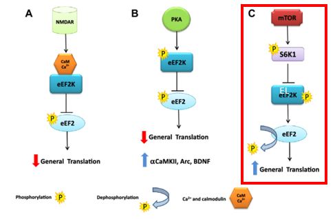
Increased gene transfer (and translation) mediated by eEF2 dephosphorylation via activation of mTORC1
In addition, high loads activated the MEK-ERK1/2-dependent signalling cascade that inhibits the TSC1/2 protein complex, which, in turn, is a Rheb protein inhibitor, the activation of which catalyses mTORC1 binding, thereby activating the entire cascade of reactions that stimulates protein synthesis and cell proliferation.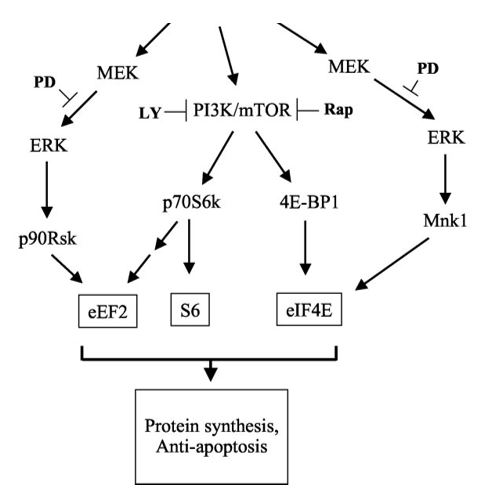
Increased protein synthesis through indirect mechanisms dependent on activation of the MEK-ERK signalling cascade
Activation of the MEK-ERK signalling cascade increases eIF4E (Mnk1 pathway) and eEF2 (p90RSK pathway) activity indirectly, whose functions have been described above and can be observed in the previous image.
There are other mechanisms that the authors point out as potential stimulators of muscle protein synthesis, such as FoxO1 dephosphorylation, which induces an increase in protein synthesis mediated by a decrease in the activation of 4EBP1, allowing a greater phosphorylation of eIF4E that stimulates protein synthesis or a decrease in the activity of Atrogin-1, an E3-linked enzyme of ubiquitin, mediator of the ubiquitin proteasome pathway; which favours proteolysis.
Summary and primary takeaway
I don’t want to conclude without noting the number of mechanisms cited that positively regulate oncogenesis and tumour cell proliferation, making the safety of stimulating these pathways dubious to say the least.
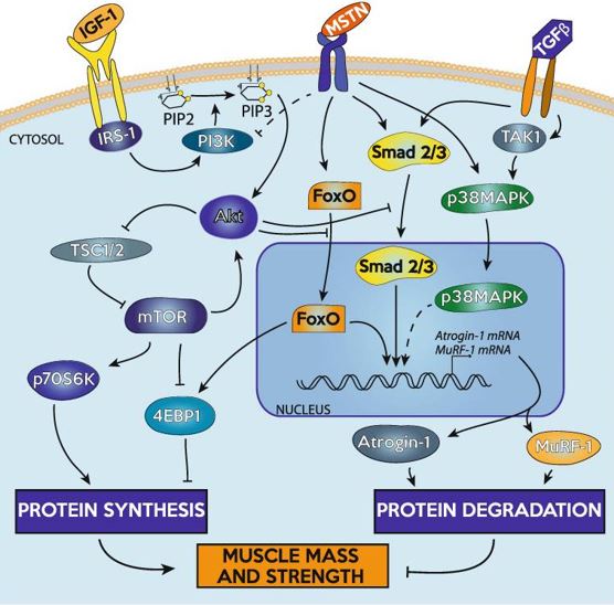
Regulation of protein turnover. Special emphasis on the activity of FoxO as an activator of 4EBP1 and Atrogin-1 and its proteolytic activity.
If you train executing 15 reps with 100kg you’ll “get” the same hypertrophy as 8 reps with 120kg
And this is due to the activation of different pathways, one dependent on mTORC1 and another on MEK-ERK 1/2
Conclusions
- Train however you like as the results will be similar
- Training volume (measured in tonnage) appears to be a completely secondary aspect versus normalised intensity at maximum volitional effort
- The more advanced you are, the more you should use both ranges to take advantage of both tracks
Bibliographic Sources
- Mitchell, C. J., Churchward-Venne, T. A., Cameron-Smith, D., & Phillips, S. M. (2015). What is the relationship between the acute muscle protein synthesis response and changes in muscle mass? Journal of Applied Physiology (Bethesda, Md. : 1985), 118(4), 495–497.
- Lysenko, E. A., Popov, D. V, Vepkhvadze, T. F., Sharova, A. P., & Vinogradova, O. L. (2019). Signaling responses to high and moderate load strength exercise in trained muscle. Physiological Reports, 7(9), e14100.
- Schoenfeld, B. J., Peterson, M. D., Ogborn, D., Contreras, B., & Sonmez, G. T. (2015). Effects of Low- vs. High-Load Resistance Training on Muscle Strength and Hypertrophy in Well-Trained Men. Journal of Strength and Conditioning Research, 29(10), 2954–2963.
Related Posts
- Intermediate Guide to Bodybuilding
- Rest-Pause: Advanced Method for Muscle Mass Gain
- Muscle fibre types and their relation to sport