 Fitness, Nutrition, Health and Sports Blog In the HSNstore Blog you will find tips about Fitness, sport in general, nutrition, and health – HSNstore.com
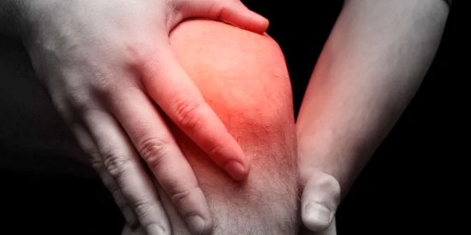

Let’s talk about the basics of knee ligament injuries: collateral, anterior and posterior cruciate; and the most suitable treatment
Knee anatomy
Knee stability is maintained by passive anatomical elements, including the ligaments.
Injury to these will show signs of laxity in different planes and, consequently, joint instability; on the other hand, active anatomical elements are mainly represented by muscles.
From this, it’s important to understand that a knee can be lax but stable or, conversely, unstable without being lax.
Functionally, the knee ligament structures can be grouped into three:
- A central pivot formed by the anterior cruciate ligament (ACL) and the posterior cruciate ligament (PCL).
- An internal compartment made up of medial structures where we find the medial collateral ligament (MCL), formed by a deep and a superficial bundle, and the posterior oblique ligament and the semimembranosus reflex tendon.
- An external compartment mainly formed by the lateral collateral ligament (LCL), and the tendons of the popliteus muscle and the biceps femoris muscle.
Collateral ligament injuries
They can occur on the lateral collateral ligament or medial collateral ligament (more common in the MCL) and can be classified as sprains or tears.
TEARS are usually accompanied by other injuries as a result of intense trauma.
Injuries during sports practice
However, in sports, sprains are more common due to forced positions, in valgus or varus, which stretch the corresponding ligament. One of the most common injuries athletes suffer in the ligaments is a sprain of the medial collateral ligament (medial collateral ligament – MCL).
This happens when the leg is blocked or receives a blow from the outside. When this occurs, the person leans on the lateral part of the knee with a forced movement, the joint moves outward (valgus) and a sprain occurs.

Treatment
- Rest, ice, compression, and elevation immediately.
- Do not put weight on the injured knee’s foot.
- For grade I and/or grade II sprains, a splint should be placed in extension until pain and swelling decrease.
Grade III sprains, on the other hand, may require surgical intervention and casting
Rehabilitation
In a later phase, some of these methods and exercises are usually recommended:
- Lymphatic drainage
- Isometric strength exercises for the knee’s extension and flexion muscles

These are easier to perform in open kinetic chain (quad extensions on machine, hamstring curls, etc.)
- Training of PNF (proprioceptive neuromuscular facilitation) with light loads.
- Mobilization of the patella, as well as the knee joint, in flexion and extension.
- Stationary bike.
- Water jogging.
- Isokinetic training for knee extension and flexion muscles in closed kinetic chain (squats, for example).
- Proprioceptive exercises for leg axis mobilization, avoiding knee rotation.

Anterior Cruciate Ligament (ACL)
The anterior cruciate ligament is the ligament that breaks most often in the knee joint.
A tear of the anterior cruciate ligament, isolated or combined with meniscal or collateral ligament injuries, causes long-term degenerative changes, especially if not properly treated.
Possible injury mechanisms
- Mechanism with knee in semiflexion, forced valgus (knee inward), and external tibial rotation: can cause an MCL injury, internal meniscal tear, and ACL tear. This set of injuries is known as the “O’Donoghue’s unhappy triad.”
- Mechanism with knee in slight flexion, forced varus (knee outward), and internal tibial rotation: which will cause an ACL injury, then an LCL injury, and internal or external meniscal tear.
- Mechanism with knee in extension and forced valgus (knee inward): which will cause an MCL injury and secondarily an ACL or PCL injury.
- Mechanism with knee in extension and forced varus (knee outward): which will cause an LCL injury and possibly an ACL injury.
- In pure frontal plane: a sudden hyperextension can cause a pure ACL injury.
In any of these five cases, it seems that the stronger the quadriceps activation during eccentric contraction, the higher the chance of ACL damage
Women are more prone to ACL injuries
Due to the wider pelvis in women, the ACL is subjected to additional pressure compared to men when any of the above mechanisms occur.

Menisci – ACL relationship
ACL insufficiency leads to meniscal injuries that alter joint movement patterns.
An example of this interdependence between different anatomical structures in the knee joint is that 80% of patients have a meniscal tear two years after tearing the ACL without proper treatment, which justifies surgical indication.
Treatment
Depending on age, type of injury, and patient activity, different options can be offered, from conservative treatment to surgery.
Rest is required, using a knee brace or splint for 3-4 weeks, with cryotherapy or ice on the knee consecutively for 72 hours. After that, start a counter-resistance program with progressive loads (starting against gravity resistance)
Rehabilitation
Isometric exercises for the quadriceps start in the first week and continue until the eighth week, accompanied by electrostimulation and stretching and hip mobility exercises.

Swimming is recommended from six weeks. Direct proprioceptive control (PNF) exercises are also very important from seven weeks.
After three months, agility exercises, lateral control, and full weight-bearing are prescribed, starting gym machine exercises.
Finally, at 5 months, running and isokinetic exercises with additional load can begin.
After ACL repair with grafts or allografts, sports should not be resumed before 7 to 8 months.

Quadriceps and hamstring muscle strength and jump distance should be 80 to 90% compared to the other leg. Ideal joint mobility is from 0º to 140º
Posterior Cruciate Ligament (PCL)
Injury mechanism
PCL tears are rare and are a typical injury in athletes, especially those who play contact sports. They are also very common in traffic accidents from impact of the flexed knee against the car dashboard.
Sprains, on the other hand, are less common and there’s not much more to add compared to previous posts
In sports, PCL injury occurs with the knee in hyperflexion and the ankle in plantar flexion, although they can also be injured by hyperextension.

Treatment
Some authors believe that conservative treatment of the PCL yields good results and surgery is not necessary in athletes who can reduce their sports activity. However, long-term results are not good.
When joint laxity is small and the PCL injury is isolated, two weeks of immobilization and unloading with crutches can be recommended
Afterwards, a program of mobility exercises and quadriceps strengthening will continue as a natural element that prevents the tibia from moving backward.

Non-surgical treatment can also be recommended for chronic PCL injuries, with an orthosis (knee brace), although it does not prevent posterior tibial displacement.

Sources
- A. Hüter-Becker. Rehabilitation in sports. Editorial Paidotribo (Barcelona) (2005).
- Keith L. Moore, Anne M. R. Agur, Marion E. Moore. Clinically oriented anatomy. 5th ed. Editorial Médica Panamericana, 2007.
- Honkamp NJ, Ranawat AS, Harner CD. Knee: Posterior cruciate ligament injuries in the adult. In: DeLee JC, Drez D Jr, Miller MD, eds. DeLee and Drez’s Orthopaedic Sports Medicine. 3rd ed. Philadelphia, Pa: Saunders Elsevier; 2009:chap 23, section E.
- Mitch Kaplan. Athlete injury guide. Ed. Hispano Europea (Barcelona) (2004).
- Roald Bahr, Sverre Maehlum. Sports injuries: Diagnosis, treatment and rehabilitation. Editorial Médica Panamericana, 2007.
- Ronald P. Pfeiffer and Brent C. Mangus. Sports injuries. Editorial Paidotribo (Barcelona) (2007).
- J Sci Orthop 7:1-5 Rossi MJ, Lubowitz JH, Guttmann D. The skier’s knee. Arthroscopy 2003 Jan; 19 (1):75-84
- William E. Garret, Donald T. Kirkendall, S. Robert Contiguglia. Soccer medicine. Editorial Paidotribo, 2005.
Related posts
- Everything you need to know about ankle sprains
- Learn how to fight shoulder pain
- What is Lymphatic Drainage?