 Fitness, Nutrition, Health and Sports Blog In the HSNstore Blog you will find tips about Fitness, sport in general, nutrition, and health – HSNstore.com

With the following article, we’ll take a closer look at the Basics of the Muscular System
At HSN, we’re going to take a step forward in learning advanced concepts.
When someone wants to design a hypertrophy training program, it’s interesting to know how the muscle works to produce force through the joints to create the movement needed to perform any exercise repetition. For this, we need to understand muscle physiology.
Knowing this, we’ll understand how muscle contraction happens, which is the key to hypertrophy.
Macrostructure of skeletal muscle
Within the macrostructure, the muscle is covered by connective tissue called epimysium.
This is made up of hundreds of muscle fascicles, which always end at both ends in what’s called a tendon that connects to the bone with a special connective tissue called periosteum, which covers all bones.
As we mentioned, the muscle has two poles or points that attach to the bones. They have different names depending on their location:
- In the limbs: proximal, if it’s the closest to the trunk, and distal
- In the trunk: superior for the one closest to the head and inferior for the one closest to the feet.
- You can also use the traditional naming: origin for the point closest to the center of the body and insertion for the point farthest from the center of the body.

Microstructure of skeletal muscle
Inside the epimysium, muscle fibers group into bundles.
These are called fascicles and are covered by a layer of connective tissue called perimysium. Inside, each fiber also has a connective tissue layer called endomysium, plus the cell membrane called sarcolemma.
If you isolate a muscle fiber, inside it just beyond its membrane, you see the cytoplasm of the muscle cell called sarcoplasm. This is made up of thousands of myofibrils.
These are what make possible what we’re after, the muscle contraction

Myofibrils
Myofibrils are basically made up of two types of myofilaments: thin actin filaments and thicker myosin filaments.
Both types of filaments are organized longitudinally forming the smallest contractile unit of skeletal muscle called the sarcomere.
M and Z lines
Myosin filaments anchor at the so-called M line in the center of the sarcomere, while actin filaments are at the ends, anchored to the Z line. These Z lines run along the entire myofibril.

Each myosin filament is surrounded by six actin filaments, and each actin filament is surrounded by three myosin filaments. This arrangement of actin and myosin filaments gives muscle its light and dark color and the striated appearance you see in bodybuilders when they have very low body fat.
Sliding filament theory
As we said, it basically involves a sliding of actin filaments in the sarcomere over the myosin filaments.
Cross-bridges
This causes the Z lines to move, which makes the muscle contract. The movement happens by a bending of the myosin cross-bridges, but to produce the full movement, many bends must happen along the whole muscle.
Calcium
A very important element, not only for bone creation, is calcium. Its release generates tension in the muscle that causes contraction.
When at rest, calcium levels in the myofibrils are very low, which means few myosin cross-bridges connect with actin filaments.
For the myosin cross-bridges to bend, they first have to attach to the actin filaments.
To make that connection, calcium is needed.
This calcium is released from the sarcoplasmic reticulum when a nerve impulse arrives, and it binds to troponin (a protein distributed along the actin filament). This binding causes a change in tropomyosin (another protein in the groove of the actin filament), allowing the myosin cross-bridge head to bind faster to the actin filament and causing the cross-bridge to bend.
ATP
For the cross-bridges to bend, energy is needed, which comes from the hydrolysis of ATP (adenosine triphosphate) into ADP (adenosine diphosphate) and phosphate, catalyzed by the myosin ATPase enzyme.
There’s a continuous need for an ATP molecule to replace the ADP so actin can dissociate from the myosin cross-bridge and keep the contraction going, as long as calcium is available to bind to troponin, which is why calcium is so important.
When all these steps happen sequentially and repeatedly along the whole muscle fiber, the muscle shortens noticeably. This will continue as long as calcium is present in the myofibril, ATP is available to dissociate actin and myosin, and myosin ATPase is present to hydrolyze ATP.
This all ends when the muscle stops being stimulated. Then, calcium returns to the sarcoplasmic reticulum, preventing actin and myosin filaments from binding. This separation puts them back in a dissociated state, which means muscle relaxation of the muscle/group in question.
Summary of the Muscle Contraction Steps
We can explain it in these 5 phases:
- Resting state. There’s little calcium and therefore few bends of the myosin cross-bridges.
- Excitation-contraction coupling phase. A stimulus releases calcium that causes more bends of the myosin cross-bridges on the actin filaments.
- Contraction. Dissociation of actin and the myosin cross-bridge by ADP release from ATP hydrolysis to generate muscle contraction.
- Recharge phase. Calcium, ATP, and myosin ATPase are needed.
- Relaxation. The muscle relaxes as myosin and actin dissociate when calcium is stored back in the sarcoplasmic reticulum.
Types of muscle fibers
It’s important to know that not all muscle fibers are the same. There are different types depending on metabolic and contractile characteristics.
They mainly group into two categories explained below:
Type I fibers
These are slow-twitch fibers.
They use a lot of oxygen. Their function is mainly based on cellular respiration. That’s why they have great fatigue resistance since they have many mitochondria, high capillary density, and high aerobic enzyme activity.
Because of their high endurance, they have less strength, mainly due to their slow contraction.
These fibers don’t have a big hypertrophy factor, they don’t grow much.
Type II
These are fast-twitch fibers.
They’re the ones that truly promote individual strength. They grow the most and are ideal for exercises like sprinting, weightlifting, fitness… The downside is they fatigue very quickly compared to the previous type.
These fibers are divided into two subtypes:
- Type IIB. These are fast glycolytic fibers. They get tired quickly and have few mitochondria, low aerobic enzyme activity, and few capillaries. They’re key for intense exercises with high contraction power.
- Type IIA. These are fast oxidative fibers. More like type I because they also use oxygen, so they last longer without getting tired than type IIB (but less than type I), and have more mitochondria, higher aerobic enzyme activity, and more capillaries than type IIB.
Types of Muscle Actions
There are 3 muscle actions we can differentiate:
- Concentric (CON),
- Isometric (ISO), and
- Eccentric (EXC).

Let’s see how each one works:
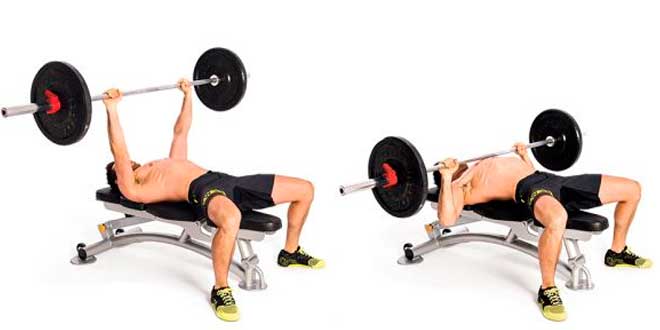
Concentric muscle actions
These involve the muscle’s main action, which is shortening.
It happens when the total tension from all the cross-bridges (see part II) exceeds the muscle’s resistance to shortening. This action brings the two ends of the bones forming a joint closer. Concentric actions are considered dynamic actions.
For example, in a biceps curl, both the cross-bridges of the biceps brachii and the elbow flexors overcome the resistance caused by the total weight (bar, plates, arm, forearm, and hand).
Isometric muscle actions
This action is also known as static.
Since there’s no movement when it happens, the muscle produces force but its length stays the same (static) and the joint angle doesn’t change. The tension from all cross-bridges equals the resistance from the total load (which is why it stays static).
For example, in a biceps curl, you stop the movement halfway, where the force from the biceps brachii and elbow flexors equals the total weight force (bar, plates, arm, forearm, and hand).
Eccentric muscle actions
Here, force is produced while the muscle lengthens.
Like concentric actions, these are dynamic actions (there’s movement). It happens when the force from the muscle’s cross-bridges is less than the resistance applied.
For example, in a biceps curl, this happens during the controlled lowering phase, where the total force from the cross-bridges slows down the movement but isn’t enough to stop it, so it’s not an isometric.
Related Posts
- Training Principles
- Training Interference Phenomenon
- Neural Load Training