 Fitness, Nutrition, Health and Sports Blog In the HSNstore Blog you will find tips about Fitness, sport in general, nutrition, and health – HSNstore.com


Saying that physical exercise is good for your health is nothing new. In a sedentary society with more and more chronic conditions every day, physical exercise is an essential tool we need to learn how to use
Exercise is Medicine
And that’s what we’re about. From the medical field, the message that “Exercise is Medicine” is sinking in, and we already have solid scientific literature backing it up, like this amazing review by Pedersen that opened our eyes to how physical exercise can prevent up to 26 chronic diseases (1).
We’ve created a mismatch or lack of coherence between what our hunter-gatherer genes expect from us and what we actually give them daily, and our health pays the price
Today’s article goes a step further. We already knew that physical exercise, especially cardiovascular (being the most studied), produced clear cardiovascular benefits that could be summed up as:
- Lowering blood pressure
- Chronic reduction of oxidative stress
- Improved functional capacity
- Increased total calorie expenditure
- Enhanced myocardial vascularization
Today we’ll focus on several lesser-known mechanisms by which exercise (both cardiovascular and strength training) produces very important cardiovascular improvements.
Antiatherogenic Effects
Continuous exposure to the hemodynamic stress caused by physical exercise leads to structural and functional adaptations in blood vessels that provide antiatherogenic properties
What is Atherogenesis?
Atherogenesis is the process by which, thanks to risk factors accumulating over the years (age, oxidative stress, insulin resistance, inflammation, LDL, etc.), an atheroma plaque (basically fat and cells) forms inside the blood vessel wall, which can eventually block it.
This 2015 meta-analysis shows how exercise intensity improves endothelial vascular function in a dose-dependent manner (3).
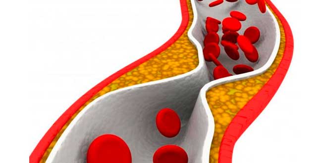
Now we have to explain why this improvement happens
Wall Stress
There’s a concept called “shear stress” in English, which could be translated as “wall stress,” illustrating the pressure blood exerts against vessel walls during exercise, triggering a series of beneficial adaptations, like increased synthesis of NO (nitric oxide), the ultimate vasodilator molecule (4).
These functional improvements are followed by structural changes, which we’ll see now.
Healthy Arteries in Trained Athletes
One of the most pioneering and curious scientific observations came from an autopsy that found Clarence DeMar, seven-time Boston Marathon champion, had “larger-than-expected coronary arteries” (6).
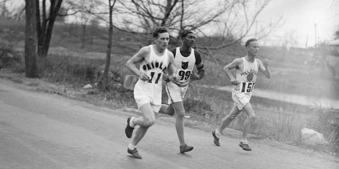
Remember, coronary arteries supply blood to the heart, our most important muscle
Later studies showed that this dilation of large vessels (aorta, subclavian, carotid) was a common trait among trained athletes, along with thinner arterial walls, all favoring a larger arterial lumen, which would give blood more room to flow smoothly, even with some atheroma plaque present (7).
Reduction of Atheroma Plaque
Also, continuing with benefits on blood vessels, the pesky atheroma plaque (which blocks blood flow and causes coronary disease, meaning you could have angina or a heart attack) has a more favorable composition in trained athletes, with increased collagen and elastin content (8). This is great because these plaques are less likely to rupture. And that’s crucial, since plaque rupture violently blocks blood flow through the vessel and causes ischemic damage.
Physical exercise also provides cardiac protection by promoting collateral vessel formation, meaning small blood vessels that form a complex network and, if a main artery gets blocked, supply some blood to the endangered tissue, minimizing damage (9).
Improvement of Autonomic Nervous System Balance
The heart is made up of muscle, valves, and a very complex nerve wiring; as you can quickly understand, the heart contracts involuntarily (imagine the stress of having to contract your own heart voluntarily)
The part of the nervous system responsible for regulating cardiac contractility (among many other functions) is called the autonomic nervous system (involuntary, independent) and is divided into two branches: sympathetic and parasympathetic.
On the other hand, many patients who die from a heart attack actually die due to a fatal arrhythmia during the infarction, causing sudden death. Ventricular fibrillation is the most common in this context and often fatal.
Heart Rate Variability
In recent years, a very interesting parameter called heart rate variability or HRV has been developed, which precisely correlates with the efferent tone of the vagus nerve (parasympathetic) to the heart (11).
High HRV means good cardiovascular health and recovery capacity.
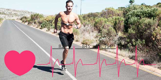
So much so that elite athletes are starting to use this marker to plan their training and recovery
Cardioprotection Against Ischemic Injuries
If you’re unlucky enough to suffer a heart attack, you’re still lucky to live in 2019…
Where most people survive thanks to a healthcare system that has advanced tremendously in recent years and allows opening the affected artery at lightning speed to save most of the damaged tissue.
We need to understand two concepts here: ischemia-reperfusion injury and cardiac preconditioning.
Reperfusion Injury
Ischemia-reperfusion injury refers to the fact that when the heart is exposed to ischemia and then reperfused thanks to medical procedures (meaning blood flows again through necrotic or pre-necrotic areas), a series of molecular events occur involving altered calcium signaling, increased oxidative damage in cells, and mitochondrial dysfunction. All this leads to myocardial damage.
Cardiac Preconditioning
On the other hand, cardiac preconditioning refers to the phenomenon where short periods of ischemia before longer arterial occlusion periods (like those in a heart attack) are cardioprotective.
Exercising regularly is buying yourself better chances of survival if you’re unlucky enough to go through this…
Creating an Anti-inflammatory Environment
Skeletal muscle is an endocrine organ that produces and releases myokines into the bloodstream, especially during muscle contraction, which exert local and systemic actions and have multiple cardiovascular benefits
The anti-inflammatory environment created by exercise protects vessels against atherosclerosis and helps stabilize previously formed atheroma plaques. For example, IL-6 released by contracting muscles increases levels of anti-inflammatory cytokines like IL-1RA or IL-10 and decreases circulating levels of pro-inflammatory ones like TNF.
Myokine Release
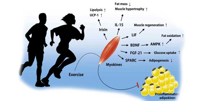
Also, myokines trigger improvements through other very different pathways:
- IL6, IL15, and FGF21 reduce muscle insulin resistance, improving glucose homeostasis.
- Irisin, METRNL, and FGF21 stimulate browning of adipose tissue, activating thermogenesis and promoting energy expenditure.
- LID, IL4, IL6, IL7, and IL15 stimulate skeletal muscle growth and maintenance, which is especially important to protect older adults against sarcopenia and visceral fat accumulation, both cardiovascular risk factors.
- Musclin (another myokine) increases mitochondrial biogenesis and cardiorespiratory fitness.
- Follistatin improves endothelial function and promotes revascularization in ischemic tissue. Irisin improves arterial relaxation and protects against endothelial damage and atherosclerosis.
- Apelin promotes vasodilation, improves myocardial contractility, and angiogenesis (14) (15) (16).
Improves Myocardial Regeneration
When you suffer a heart attack, the damaged part of the heart undergoes remodeling that is usually negative for overall heart health, facilitating heart failure and causing mortality in the first year after the infarction to remain high
Exercise has been shown to mitigate this adverse myocardial remodeling that occurs after ischemic damage, for example by reducing left ventricular end-systolic volume, a strong predictor of mortality after myocardial infarction (17).
Also, exercise directly stimulates a series of stem cells called “circulating angiogenic cells”, whose levels inversely relate to cardiovascular disease (18).
Prevents Sarcopenia
I’ll be brief: muscle strength is inversely related to cardiovascular disease development, mortality from it, and all-cause mortality in kids, teens, and adults (20)
Good muscle mass protects against cardiovascular events in the elderly, and forearm circumference is a good survival predictor in those over 70 (21). Strength, in a word, is health.
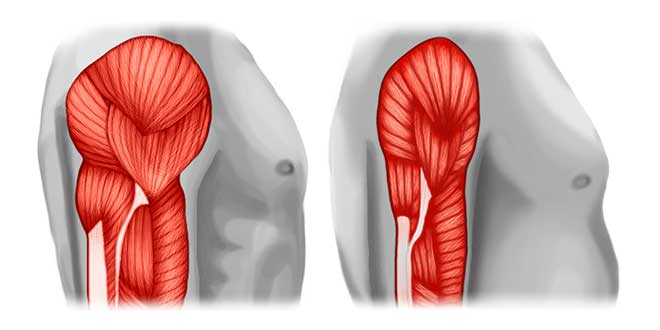
Sarcopenia is another big epidemic nowadays, causing many premature deaths
Dangers of Sarcopenia
The pathophysiological mechanisms that make sarcopenia dangerous are:
- Systemic inflammation, partly due to altered myokine secretion, which as we said reduces NO availability in vessels and thus vasodilation.
- Insulin resistance, linked to worse glucose control, which in turn relates to mitochondrial and endothelial damage and left ventricular hypertrophy (22).
- Lower carnitine release from muscle, a protein that transports long-chain fatty acids to mitochondria for oxidation.
- Dysregulation in adipokine secretion
- Higher risk of disability and lower functional capacity.

The antidote is probably already on your mind: strength training
Improves Microbiota
The digestive microbiota, that great unknown seemingly involved in every disease imaginable…
It seems that high cardiorespiratory fitness, an active lifestyle, and regular physical exercise can modulate the microbiota through the following mechanisms:
- Increased bacterial diversity, a parameter associated with better health and disease prevention.
- Higher fecal concentrations of short-chain fatty acids like butyrate, an indicator of gut health.
- Greater proportion of healthy species, like Faecalibacterium prausnitzii, Roseburia hominis, and Akkermansia muciniphila, and better levels of those linked to metabolic disease and obesity (e.g., Clostridium coccoidea).
Conclusions
We already knew physical exercise is beneficial…
But day by day, the scientific community works hard to investigate new pathways and mechanisms through which exercise produces these positive effects.

We need to stop thinking of physical exercise as just a pastime for our free time and start seeing it as powerful medicine and, of course, a developing science
References
- Pedersen B, Saltin B. Exercise as medicine–evidence for prescribing exercise as therapy in 26 different chronic diseases. J Med Sci Sport. 2015;
- Fiuza-Luces C, Santos-Lozano A, Joyner M, Carrera-Bastos P, Picazo O, Zugaza JL, et al. Exercise benefits in cardiovascular disease: beyond attenuation of traditional risk factors. Nature Reviews Cardiology. 2018.
- Ashor AW, Lara J, Siervo M, Celis-Morales C, Oggioni C, Jakovljevic DG, et al. Exercise Modalities and Endothelial Function: A Systematic Review and Dose–Response Meta-Analysis of Randomized Controlled Trials. Sports Medicine. 2015.
- Erkens R, Suvorava T, Kramer CM, Diederich LD, Kelm M, Cortese-Krott MM. Modulation of Local and Systemic Heterocellular Communication by Mechanical Forces: A Role of Endothelial Nitric Oxide Synthase. Antioxid Redox Signal. 2017;
- Hambrecht R, Wolf A, Gielen S, Linke A, Hofer J, Erbs S, et al. Effect of Exercise on Coronary Endothelial Function in Patients with Coronary Artery Disease. N Engl J Med. 2000;
- Dill DB. Marathoner demar: Physiological studies. J Natl Cancer Inst. 1965;
- Haskell WL, Sims C, Myll J, Bortz WM, St. Goar FG, Alderman EL. Coronary artery size and dilating capacity in ultradistance runners. Circulation. 1993;
- Shimada K, Mikami Y, Murayama T, Yokode M, Fujita M, Kita T, et al. Atherosclerotic plaques induced by marble-burying behavior are stabilized by exercise training in experimental atherosclerosis. Int J Cardiol. 2011;
- Heaps CL, Robles JC, Sarin V, Mattox ML, Parker JL. Exercise training-induced adaptations in mediators of sustained endothelium-dependent coronary artery relaxation in a porcine model of ischemic heart disease. Microcirculation. 2014;
- Bhella PS, Hastings JL, Fujimoto N, Shibata S, Carrick-Ranson G, Palmer MD, et al. Impact of lifelong exercise “dose” on left ventricular compliance and distensibility. J Am Coll Cardiol. 2014;
- Billman GE, Csepe T, Fedorov V V., Cagnoli KL, Wright P, Mohler PJ, et al. Exercise training-induced bradycardia: evidence for enhanced parasympathetic regulation without changes in intrinsic sinoatrial node function. J Appl Physiol. 2015;
- Billman GE. Cardiac autonomic neural remodeling and susceptibility to sudden cardiac death: effect of endurance exercise training. Am J Physiol Circ Physiol. 2009;
- Thijssen DHJ, Redington A, George KP, Hopman MTE, Jones H. Association of exercise preconditioning with immediate cardioprotection: A review. JAMA Cardiology. 2018.
- Subbotina E, Sierra A, Zhu Z, Gao Z, Koganti SRK, Reyes S, et al. Musclin is an activity-stimulated myokine that enhances physical endurance. Proc Natl Acad Sci. 2015;
- Medina-Inojosa JR, Somers VK, Thomas RJ, Jean N, Jenkins SM, Gomez-Ibarra MA, et al. Association between adiposity and lean mass with long-term cardiovascular events in patients with coronary artery disease: No paradox. J Am Heart Assoc. 2018;
- Drevon CA, Raastad T, Thiede B, Haugen F, Norheim F, Rustan AC. Proteomic identification of secreted proteins from human skeletal muscle cells and expression in response to strength training. Am J Physiol Metab. 2011;
- Haykowsky M, Scott J, Esch B, Schopflocher D, Myers J, Paterson I, et al. A Meta-analysis of the effects of Exercise Training on Left Ventricular Remodeling Following Myocardial Infarction: Start early and go longer for greatest exercise benefits on remodeling. Trials. 2011;
- Zalos G, Finkel T, Hill JM, Schenke WH, Waclawiw MA, Halcox JPJ, et al. Circulating Endothelial Progenitor Cells, Vascular Function, and Cardiovascular Risk. Obstet Gynecol Surv. 2004;
- Vasa M, Fichtlscherer S, Adler K, Aicher A, Martin H, Zeiher AM, et al. Increase in circulating endothelial progenitor cells by statin therapy in patients with stable coronary artery disease. Circulation. 2001;
- Celis-Morales CA, Welsh P, Lyall DM, Steell L, Petermann F, Anderson J, et al. Associations of grip strength with cardiovascular, respiratory, and cancer outcomes and all cause mortality: Prospective cohort study of half a million UK Biobank participants. BMJ. 2018;
- Kamiya K, Masuda T, Matsue Y, Hamazaki N, Matsuzawa R, Tanaka S, et al. Prognostic Usefulness of Arm and Calf Circumference in Patients ≥65 Years of Age With Cardiovascular Disease. Am J Cardiol. 2017;
- Kinugasa Y, Yamamoto K. The challenge of frailty and sarcopenia in heart failure with preserved ejection fraction. Heart. 2017;
- Serra-Rexach JA, Bustamante-Ara N, Hierro Villarán M, González Gil P, Sanz Ibáñez MJ, Blanco Sanz N, et al. Short-term, light- to moderate-intensity exercise training improves leg muscle strength in the oldest old: A randomized controlled trial. J Am Geriatr Soc. 2011;
Related Posts
- Cardiovascular health – How to take care of our heart: foods and good routines
- Cardiovascular Exercises: Improve your fitness and burn fat
- Benefits of aerobic exercise
- What to eat before exercising?